42 external structure of the heart with labels
Ch. 19 Circulatory System- heart Flashcards | Quizlet Correctly label the external anatomy of the anterior heart. Place the labels in order denoting the flow of blood through the pulmonary circuit beginning with the right atrium and ending in the left atrioventricular valve. The first and last structures are given. Right atrium 1. tricuspid valve 2. right ventricle 3. pulmonary valve Heart Anatomy: Heart Dissection The major vessels of the heart are found at the base of the heart, along with the upper chambers, the right atrium (C) and left atrium (D). The atria are collapsed, but in a functioning heart, they would be stretched full of blood. The majority of the heart tissue consists of the ventricles. The left ventricle (F) is stiff and solid because it ...
Structure of the Heart | SEER Training The human heart is a four-chambered muscular organ, shaped and sized roughly like a man's closed fist with two-thirds of the mass to the left of midline. The heart is enclosed in a pericardial sac that is lined with the parietal layers of a serous membrane. The visceral layer of the serous membrane forms the epicardium. Layers of the Heart Wall

External structure of the heart with labels
Heart Anatomy: size, location, coverings and layers : Anatomy & Physiology Heart Anatomy. The heart is around the size of a fist and weighs between 250-350 grams (less than a pound). Enclosed within the mediastinum, the medial cavity of the thorax, the heart extends obliquely from the second rib to the fifth intercostal space. It rests on the superior surface of the diaphragm, lies posterior to the sternum and ... Heart Anatomy | Anatomy and Physiology | Course Hero The wall of the heart is composed of three layers of unequal thickness. From superficial to deep, these are the epicardium, the myocardium, and the endocardium. The outermost layer of the wall of the heart is also the innermost layer of the pericardium, the epicardium, or the visceral pericardium discussed earlier. Figure 6. The Anatomy of the Heart, Its Structures, and Functions Updated on April 05, 2020. The heart is the organ that helps supply blood and oxygen to all parts of the body. It is divided by a partition (or septum) into two halves. The halves are, in turn, divided into four chambers. The heart is situated within the chest cavity and surrounded by a fluid-filled sac called the pericardium.
External structure of the heart with labels. Heart Anatomy Labeling Game This is an online quiz called Heart Anatomy Labeling Game There is a printable worksheet available for download here so you can take the quiz with pen and paper. Your Skills & Rank Total Points 0 Get started! Today's Rank -- 0 Today 's Points One of us! Game Points 19 You need to get 100% to score the 19 points available Actions Solved Art-Labeling Activity: Overview of the external - Chegg art-labeling activity: overview of the external anatomy of the heart anterior view res great cardiac vein aortic arch right coronary artery left coronary artery left pulmonary veins ascending aorta left pulmonary artery anterior interventricular artery superior vena cava pulmonary trunk auricle of left atrium circumflex artery auricle of right … Correctly Label The Following External Anatomy Of The Anterior Heart ... The right atrium, the anterior descending aorta, and the vasculature are all essential organs of the cardiovascular system. The external anatomy of the human heart consists of the four chambers that form the apex of the heart. Each chamber has an apex that corresponds to a box. There are two boxes on each side of the heart: the atria and the ... Chapter 22 Heart Flashcards | Quizlet Label the coronary arteries in an anterior view of the heart. Label the order that blood flows through in the heart, using the arrows as guides. Label the components of the heart wall. Label the components of the heart as seen from a posterior view. Label the major coronary veins. Label the components of the conduction system.
Heart Anatomy: Labeled Diagram, Structures, Function, and Blood Flow Let's begin with the chambers of the heart. There are 4 chambers, labeled 1-4 on the diagram below. To help simplify things, we can convert the heart into a square. We will then divide that square into 4 different boxes which will represent the 4 chambers of the heart. BYJUS BYJUS Lesson | The Heart - External Structure | Encounter Edu In this lesson students begin their exploration of the circulatory system, labelling a diagram of the external structures and identifying arteries and veins. They will go on to explain where blood enters and leaves the heart. Learning outcomes Surface projections of the heart: Borders and landmarks - Kenhub 1/4. The surface projections of the heart represent points on the thoracic wall that map out the outline and valves of the heart. These include four borders (superior, right, inferior, left) and four valves (left atrioventricular, right atrioventricular, aortic, pulmonary). The main reference points used for the surface projections of the heart ...
How to Draw the Internal Structure of the Heart (with Pictures) To finish drawing the aorta, draw three nubs at the top of the loop. After you draw these, erase the lines connecting from one side of the bottom of the nub to the other. Add tilted circles to the top of all of the nubs. Draw a circle at the bottom of the aorta, adjacent to the left ventricle. Heart - External Features - Anatomy QA Apex beat. Is the lowermost and outermost thrust of the heart, felt on the front of the chest. In adults it is felt in the left 5 th intercostal space 9cm. from the median plane (just medial to the midclavicular line). In infants it is felt in the 3 rd intercostal space just lateral to the midclavicular line.. Dextrocardia. It is a congenital anomaly in which the heart lies on the right side ... The Heart - Science Quiz - GeoGuessr This science quiz game will help you identify the parts of the human heart with ease. Blood comes in through veins and exists via arteries—to control the direction of the flow, the heart has four sets of valves. The heart is an amazing machine with a lot of moving parts—let this quiz game help you find your way around this most vital of organs. Label the Heart - The Biology Corner Shows a picture of a heart with letters and blanks for practice with labeling the parts of the heart and tracing the flow of blood within the heart.
Biology 156
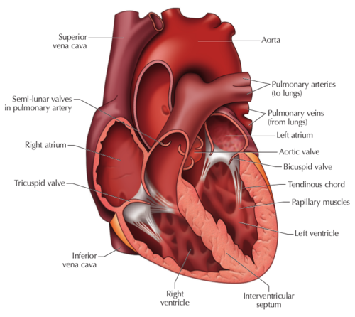
A Labeled Diagram of the Human Heart You Really Need to See The human heart, comprises four chambers: right atrium, left atrium, right ventricle and left ventricle. The two upper chambers are called the left and the right atria, and the two lower chambers are known as the left and the right ventricles. The two atria and ventricles are separated from each other by a muscle wall called 'septum'.
Virtual Squid Dissection
Structure Of The Heart | A-Level Biology Revision Notes The heart is a hollow muscular organ that lies in the middle of the chest cavity. It is enclosed in the pericardium, which protects the heart and facilitates its pumping action. The heart is divided into four chambers: The two atria (auricles): these are the upper two chambers. They have thin walls which receive blood from veins.
Natural Sciences Grade 9
Human Heart - Diagram and Anatomy of the Heart - Innerbody Because the heart points to the left, about 2/3 of the heart's mass is found on the left side of the body and the other 1/3 is on the right. Anatomy of the Heart Pericardium. The heart sits within a fluid-filled cavity called the pericardial cavity. The walls and lining of the pericardial cavity are a special membrane known as the pericardium.
Post a Comment for "42 external structure of the heart with labels"