43 microscope images with labels
Microscope- Definition, Parts, Functions, Types, Diagram, Uses It is a type of fluorescence microscope that is used to produce 2-D or 3-D images of relatively thick specimens. In this type, the excitation light is focused on a specific spot of sample lying on the focal plane. The focus spot is optically manipulated to scan the entire sample and generate a 3-D image. Electron Microscopy Images - Dartmouth We have a library of images recorded using our scanning and transmission electron microscopes. Some are shown below and others elsewhere. These images are in the public domain. If you have questions about the images or want some specific images contact Max Guinel . Hibiscus Flower (August 2021) Morphy Amorphophallus titanum anther cross section.
Label-free image-encoded microfluidic cell sorter with a scanning ... The microfluidic-based, label-free image-guided cell sorter offers a low-cost, high information content, and disposable solution that overcomes many limitations in conventional cell sorters. ... The first experiment demonstrated a sorting purity of 97%, verified by 233 microscope images; the second experiment demonstrated 100% sorting purity ...
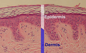
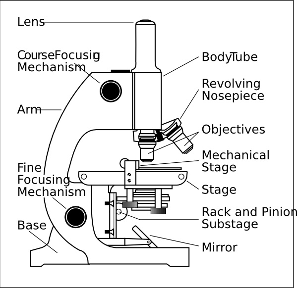
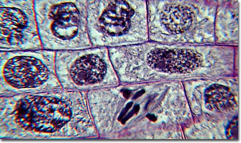
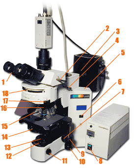
Microscope images with labels
Microscope Types (with labeled diagrams) and Functions This is an advanced microscope that has specific application in viewing, observing and measuring the optical thickness and phase of completely transparent specimens and objects. A tiny interferometer is used and a specimen is placed on beam path of it. This path is split and then rejoined to create two superimposed images of the specimen in focus. Microscope Parts, Function, & Labeled Diagram - slidingmotion Microscope parts labeled diagram gives us all the information about its parts and their position in the microscope. Microscope Parts Labeled Diagram The principle of the Microscope gives you an exact reason to use it. It works on the 3 principles. Magnification Resolving Power Numerical Aperture. Parts of Microscope Head Base Arm Eyepiece Lens Mushroom Microscope Images, Stock Photos & Vectors | Shutterstock 1,335 mushroom microscope stock photos, vectors, and illustrations are available royalty-free. See mushroom microscope stock video clips. of 14. paint under microscope microscope food electron microscope images mold microscope mycelium bacteria microscope. Try these curated collections. Search for "mushroom microscope" in these categories. Next ...
Microscope images with labels. 50 Striking Microscopic Images of Viruses and Bacteria 50 Striking Microscopic Images of Viruses and Bacteria March 07, 2022 1/50 Colorized transmission electron micrograph showing H1N1 influenza virus particles. Surface proteins on the virus particles... Can You Correctly Label These Images Of Chromosomes Labels can be used once more than once or not at all. Dividing human cells can be photographed during prophase and metaphase and all the 46 chromosome doublets can be arranged into 23 homologous pairs. Part A Drag the terms to their correct locations on the figure below. If a cell loses control of the cell cycle cancer can develop. Compound Microscope - Diagram (Parts labelled), Principle and Uses Compound Microscope - Diagram (Parts labelled), Principle and Uses As the name suggests, a compound microscope uses a combination of lenses coupled with an artificial light source to magnify an object at various zoom levels to study the object. A compound microscope: Is used to view samples that are not visible to the naked eye Types and parts of microscopes - Kenhub Electron microscope. The lenses condense the electron beams prior to them making contact with the specimen; which is held in place by a specimen holder, which is analogous to the mechanical stage of the light microscope.Objective lens then collects the transmitted electron beans and magnifies the image before projecting it to the fluorescent screen. ...
The Best Photos Taken Through Microscopes Will Blow You Away Since 1974, Nikon has held a photography competition to recognize excellent images taken with the assistance of a microscope. In 2021, the competition received almost 2,000 entries from 88 countries. In these images, art and science come together in a surprising and beautiful way. We looked through this year's winning images to share our favorites. Researchers demonstrate label-free super-resolution microscopy A newly developed sub-diffraction-limit microscopy approach doesn't require fluorescent labels. The video shows the process of the data evaluation algorithm, retrieving the positions and sizes of... Plant Cell Under Microscope Labeled 40X : Young Root 2 Of Broad Bean ... Plant Cell Under Microscope Labeled 40X : ... Record the microscope images using labelled diagrams or produce digital images. Cell Structure from Make sure your straight labelling lines match the label exactly! Pink plant cells under microscope. A cell is a very tiny structure which exists in living bodies. Sperm Under Microscope with Labeled Diagram Sperm Under Microscope 400X Labeled Diagram Before that, you may also read the below-mentioned article to get a full idea of the structure of seminiferous tubules - Histological features of the seminiferous tubules with the labeled diagram Okay, first, let's see the different histological features of the seminiferous tubules of an animal.
Parts of the Microscope with Labeling (also Free Printouts) Microscopes are specially created to magnify the image of the subject being studied. This exercise is created to be used in homes and schools. the microscope layout, including the blank and answered versions are available as pdf downloads. Click to Download : Label the Parts of the Microscope (A4) PDF print version. Parts of a microscope with functions and labeled diagram Optical parts of a microscope and their functions The optical parts of the microscope are used to view, magnify, and produce an image from a specimen placed on a slide. These parts include: Eyepiece - also known as the ocular. This is the part used to look through the microscope. Its found at the top of the microscope. Cecum Histology Slide with Labeled Image and Diagram The tunica muscular layer of the provided cecum labeled image shows two distinct smooth muscle layers - inner longitudinal or oblique bundles and outer wavy bundles. Again, the cecum images show some elastic fibers in this layer. In addition, the cecum labeled image shows a thin and loose connective tissue layer with numerous blood vessels. Microscopic Morphology - BIO 2410: Microbiology - Baker College What are the shapes of the bacteria labeled 1 and 3 in this image? Shape 5A A1 Bacillus (streptobacillus) A3 Bacillus . Shape 6 1. What is the shape of the bacteria labeled in this image? ... The microscope images in this section show different bacterial structures visible using the light microscope. All images were photographed at 1000x ...
Microscope, Microscope Parts, Labeled Diagram, and Functions Microscope, Microscope Parts, Labeled Diagram, and Functions What is Microscope? A microscope is a laboratory instrument used to examine objects that are too small to be seen by the naked eye. It is derived from Ancient Greek words and composed of mikrós, "small" and skopeîn,"to look" or "see".
Simple Microscope - Parts, Functions, Diagram and Labelling Simple Microscope - Parts, Functions, Diagram and Labelling A microscope is one of the commonly used equipment in a laboratory setting. A microscope is an optical instrument used to magnify an image of a tiny object; objects that are not visible to the human eyes. Table of Contents The common types of microscopes are: What is a Simple microscope?
Microscope Parts | A Guide on their Location and Function The image of a compound microscope with labeled parts. Eyepiece It is the part that you encounter when viewing an object in the microscope from the top. This is the first lens that helps to magnify the image. Based on the magnification power, the lens can be of 5X, 10X, 15X, or more.
Compound Microscope- Definition, Labeled Diagram, Principle, Parts, Uses In order to ascertain the total magnification when viewing an image with a compound light microscope, take the power of the objective lens which is at 4x, 10x or 40x and multiply it by the power of the eyepiece which is typically 10x. Therefore, a 10x eyepiece used with a 40X objective lens will produce a magnification of 400X.
animal cell microscope labeled - Numbers Fullerton Plant cells and animal cells share some common features as both are eukaryotic cells. 560 x 364 pixel electron microscope image animal cell and organelles labeled animal cell plasma membrane organelles. So lets begin by drawing a rough-oval shape. They are different from plant cells in that they do contain cell walls and chloroplast.
Compound Microscope Parts, Diagram Definition, Application, Working ... A compound microscope can magnify the image of a tiny object up to 1000. The term compound means "multiple" or "complex". The compound microscopes is consists of two lenses includes, the objective lens (typically 4x, 10x, 40x or 100x) in a rotating nosepiece closer to the specimen, and the eyepiece lens (typically 10x) in the binocular ...
Plant Cell Under Microscope 40X Labeled : 1 - Chloroplast and cell wall ... The different images below were taken with two different types of microscopes. 1.can only turn fine adjustment 2.draw one row of cells across the middle 3.label the chloroplasts and cell wall. When using the microscope always start by focusing under low power and working your way up to high power.
Microscopy - Wikipedia A huge selection of microscopy techniques are available to increase contrast or label a sample. Four examples of transillumination techniques used to generate contrast in a sample of tissue paper. 1.559 μm/pixel. Bright field illumination, sample contrast comes from absorbance of light in the sample
Parts of a Compound Microscope and Their Functions The main parts of compound microscope are the condenser lens, the objective lens, and the eyepiece lens, and these instruments are referred to as compound microscopes. Each of these components is made up of microscope lens combinations that are required to produce magnified images with minimal artefacts and aberrations.
Amoeba Under Microscope 40X Labeled / Microscope / This is a complex ... In this lab, you will not use the . Scanning (4x), low (10x), high (40x), and oil immersion (100x). Microscope image guessing game with scanning electron microscope pictures. Labeled Euglena Under Microscope 400x - Micropedia from i.ytimg.com Cells and microscopes, cells amoeba microscopes cronodon. Also visible are hundreds of vesicles inside ...
Mushroom Microscope Images, Stock Photos & Vectors | Shutterstock 1,335 mushroom microscope stock photos, vectors, and illustrations are available royalty-free. See mushroom microscope stock video clips. of 14. paint under microscope microscope food electron microscope images mold microscope mycelium bacteria microscope. Try these curated collections. Search for "mushroom microscope" in these categories. Next ...
Microscope Parts, Function, & Labeled Diagram - slidingmotion Microscope parts labeled diagram gives us all the information about its parts and their position in the microscope. Microscope Parts Labeled Diagram The principle of the Microscope gives you an exact reason to use it. It works on the 3 principles. Magnification Resolving Power Numerical Aperture. Parts of Microscope Head Base Arm Eyepiece Lens
Microscope Types (with labeled diagrams) and Functions This is an advanced microscope that has specific application in viewing, observing and measuring the optical thickness and phase of completely transparent specimens and objects. A tiny interferometer is used and a specimen is placed on beam path of it. This path is split and then rejoined to create two superimposed images of the specimen in focus.


Post a Comment for "43 microscope images with labels"