44 chlamydomonas diagram with labels
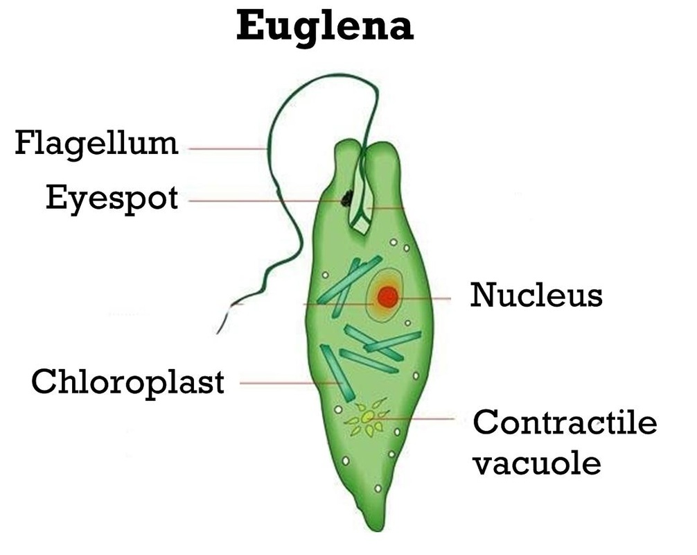
Biology Paper 1 Questions and Answers - MECS Cluster Joint Mock Exams ... On the diagram indicate the direction of movement of a nerve impulse along the neuron (1 mark) In an experiment, it was observed that when maggots are exposed to light, they move to dark areas. On the other hand, Euglena and Chlamydomonas move towards light. Name the type of response exhibited by the organisms (1mark) Blogger - Hastanta Herdian I tell you about how can we draw labelled diagram of chlamydomonas in . Draw a labelled diagram of chlamydomonas. The Unicellular Green Alga Chlamydomonas Reinhardtii As An Experimental System To Study Chloroplast Rna Metabolism Semantic Scholar from d3i71xaburhd42.cloudfront.net Chlamydomonas genes with homologs in other. Well labelled diagram ...
Diatom - Wikipedia Diatom (Neo-Latin diatoma) refers to any member of a large group comprising several genera of algae, specifically microalgae, found in the oceans, waterways and soils of the world.Living diatoms make up a significant portion of the Earth's biomass: they generate about 20 to 50 percent of the oxygen produced on the planet each year, take in over 6.7 billion metric tons of silicon each year from ...
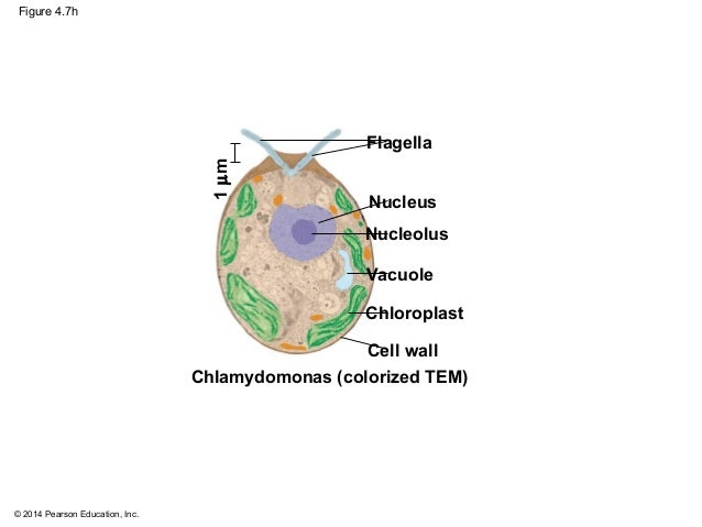
Chlamydomonas diagram with labels
Animal Cell Under The Electron Microscope - ボード「STEM: Cells ... Chlamydomonas reinhardtii, a single celled green algae, as seen under the transmission electron microscope. Now the first thing to point out when looking at images under an electron microscope is the scale. ... The diagram is very clear, and labeled here is an electron micrograph of an animal cell with the labels superimposed: At approximately ... Cnidaria: Definition, Characteristics, Examples - Biology Learner Figure: Labeled diagram of Obelia sp. General Characters of Cnidaria Their bodies are multicellular and radially symmetrical. The animals are diploblastic. The body has two layers called ectoderm and endoderm. Like jelly, mesoglea consists of two layers. Inside the body is the gastrovascular cavity, called the coelenteron. WAEC GCE Biology Questions and Answers 2021/2022 ( Essay and ... - Bekeking The diagram below illustrates a part of human skeleton. Study it and answer questions 8 to 10. 8. The diagram represent the bones of the (a) upper arm (b) lower arm (c) upper leg (d) lower leg. 9. Which of the labeled parts articulates with the head of the trochlea to form a hinge point? (a) I (b) II (c) III (d) IV. 10.
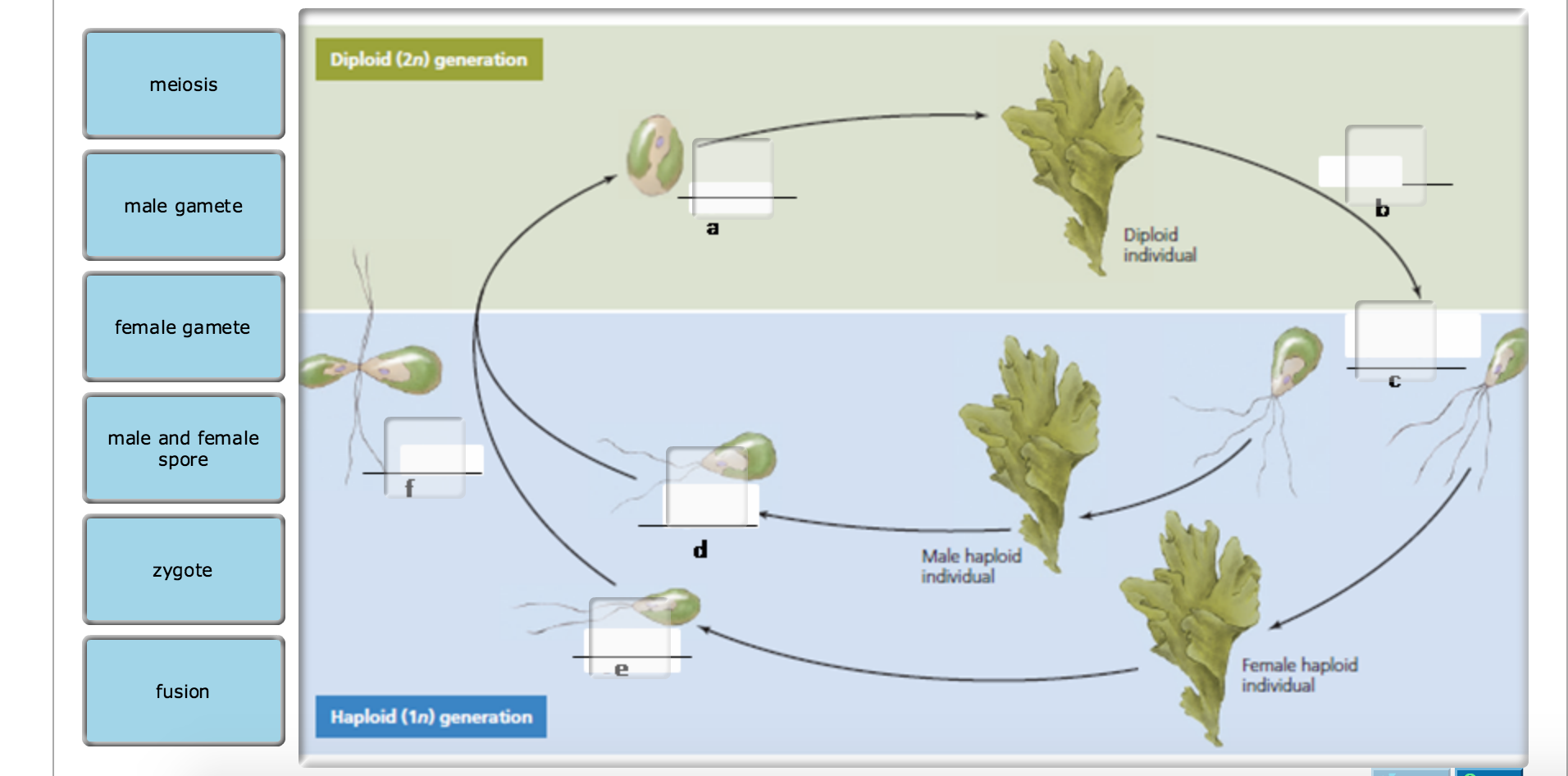
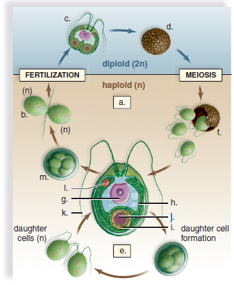
Chlamydomonas diagram with labels. Structure and Function of a Typical Bacterial Cell with Diagram August 14, 2021. Bacteria are unicellular. Their structure is a very simple type. Bacteria are prokaryotes because they do not have a well-formed nucleus. A typical bacterial cell is structurally very similar to a plant cell. The cell structure of a bacterial cell consists of a complex membrane and membrane-bound protoplast. Augmented CO2 tolerance by expressing a single H+-pump enables ... - Nature The updated Chlamydomonas reinhardtii genome and gene annotation for the RNA sequencing and DNA insertion analysis were downloaded from JGI Phytozome v13, Chlamydomonas reinhardtii v5.6 database ... › algae › morphology-ofMorphology of Chlamydomonas (With Diagram) | Algae ADVERTISEMENTS: In this article we will discuss about the external morphology of chlamydomonas. Also learn about its Neuromotor Apparatus, Electron Micrograph, Palmella-Stage with suitable diagram. 1. The organism is an unicellular alga (Fig. 11). 2. The thallus is spherical to oblong in shape but some species are pyriform or ovoid. ADVERTISEMENTS: 3. The cell is […] › algae › life-cycle-ofLife Cycle of Chlamydomonas (With Diagram) - Biology Discussion Each daughter cell develops cell wall, flagella and transforms into zoospore (Fig. 6). The zoospores are liberated from the parent cell or zoosporangium by gelatinization or rupture of the cell wall. The zoospores are identical to the parent cell in structure but smaller in size. The zoospores simply enlarge to become mature Chlamydomonas.
Flagella- Definition, Structure, Types, Arrangement, Functions, Examples The most commonly studied flagellum is the flagellum occurring in Chlamydomonas reinhardtii as a result of its size. Even though most flagella occur at the polar ends of the cells, their number and position differ as their composition and functions remain the same within a species. Created with BioRender.com Structure of Flagellum Cell- Structure and Function Archives - Biology Discussion 47 Ans. Which organelle is known as "power house" of the cell? 0 Ans. Describe prophase of mitosis. 0 Ans. How do you appreciate about the organization of cell in the living body? 1 Ans. When a tadpole turns into a frog, its tail shrinks and is reabsorbed. Is this an example of necrosis or apoptosis? Schematic Diagrams and Service Manuals - Click to Cure Cancer Schematic diagram for the measurement of the distance between gold particles and rosette TC (right) is also shown (green rosette, pink primary antibody to cellulose synthase, blue secondary antibody, red gold particle the 93 kDa antibody-labeled particles. (a) Schematic diagram for the measurement of the distance between gold particles and ... Maharashtra Board Class 11 Biology Solutions Chapter 3 Kingdom Plantae Chlamydomonas is microscopic whereas Sargassum is macroscopic; both are algae. ... Draw neat labelled diagrams. Question 1. Draw neat and labelled diagram of: (A) Spirogyra (B) Chlamydomonas Answer: ... Draw and label one example of each class. Answer: Two classes of Angiosperms are Dicotyledonae and Monocotyledonae.
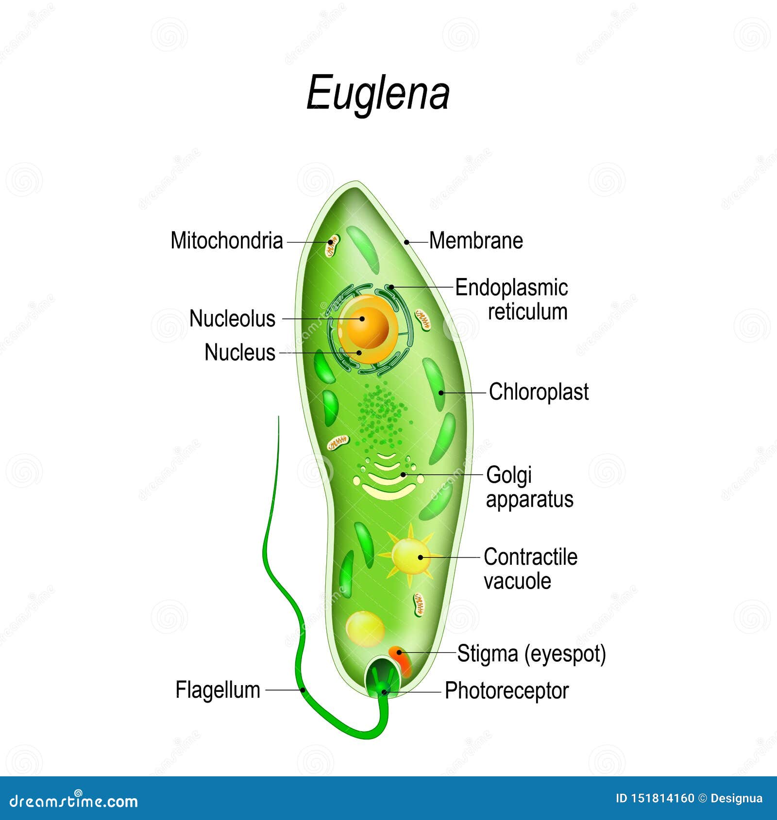
› organism › chlamydomonasStructure of Chlamydomonas (With Diagram) | Genetics In this article we will discuss about the structure of chlamydomonas (explained with labelled diagram). The unicellular green alga Chlamydomonas is haploid with a single nucleus, a chloroplast and several mitochondria (Fig. 9.3). It can reproduce asexually as well as sexually by fusion of gametes of opposite mating types (mt + and mt –). The mating type is controlled by a single nuclear gene. › biology › chlamydomonasChlamydomonas - Meaning, Structure, Life Cycle, Function and FAQs Given below is the Chlamydomonas structure with labels. The Life Cycle of Chlamydomonas . Chlamydomonas Reproduction is both sexual as well as asexual reproduction. Asexual reproduction takes place by following methods: 1. Zoospore Formation: The protoplast separates from the cell wall as it contracts. The parent cell loses its flagella, or in certain Chlamydomonas species, the flagella are absorbed. Flagella: Definition, Structure & Functions - Study.com The eukaryotic flagellum is a long, rod-like structure that is surrounded by an extension of the cell membrane like a sheath. The bulk of the structure is a filament called an axoneme. Necessary ... Animal Cells: Labelled Diagram, Definitions, and Structure Cilia are small, wiggling arm-like structures, whereas flagella are like a tail. Both structures are made of long protein fibers called microtubules, with a structure where nine microtubules form a ring around two central microtubules. Cell Membrane The cell membrane encloses the cell's contents. It monitors what comes in, and what goes out.
Overexpressing CrePAPS Polyadenylate Activity Enhances Protein ... Generation and confirmation of CrePAPS -overexpressing Chlamydomonas reinhardtii transgenic lines. ( a) The CrePAPS gene structure and the CIB1 cassette insertion position. ( b) Schematic diagram of the pJ1DCF-CrePAPS vector. Promoters and other components are labelled.
Brigitte Zimmer a typical animal cell diagram labeled black and white anatomical heart drawing with labels animal cell and plant cell diagram in hindi ... draw a human eye and label it draw the diagram of chlamydomonas draw the structure of human digestive system drawing color human digestive system
(PDF) The Dunaliella salina organelle genomes: Large sequences ... Venn diagram comparing the gene repertoires of chlamydomonadalean mitochondrial genomes. Chlamydomonadalean algae are labeled as follows: Ce = Chlamydomonas eugametos; Ch = Chlorogonium elongatum ...
› algae › structure-ofStructure of Chlamydomonas (With Diagram) | Chlorophyta In this article we will discuss about the structure of chlamydomonas with the help of suitable diagrams. Chlamydomonas is unicellular, motile green algae. The thallus is represented by a single cell. It is about 20 p,-30|i in length and 20 µ in diameter. The shape of thallus can be oval, spherical, oblong, ellipsoidal or pyriform.
Dynamic light‐ and acetate‐dependent ... - Wiley Online Library Chlamydomonas can grow photoautotrophically with just light as an energy source for carbon dioxide fixation, as well as heterotrophically with acetate as carbon source for respiration. In addition, Chlamydomonas is often grown in a mixture between both conditions (acetate and light), which is named mixotrophic growth (Hoober, 1989).
Draw neat and labelled diagram of Nephrolepis. - Sarthaks Draw neat and labelled diagram of: (A) Spirogyra (B) Chlamydomonas. asked Dec 15, 2021 in Biology by Pramsu (33.8k points) kingdom plantae; class-11; 0 votes. 1 answer. Explain in brief the two classes of Angiosperms? Draw and label one example of each class. asked Dec 15, 2021 in Biology by Pramsu (33.8k points) kingdom plantae; class-11;
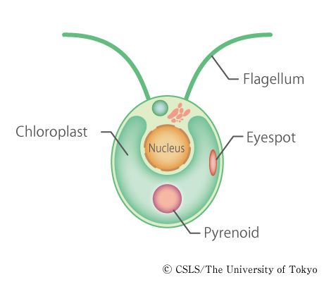
Schematic diagram of chlamydomonas | Algae - unclassified | Science textbook, Diagram, Green algae
Explain With Diagram. Chlamydomonas is a unicellular, motile freshwater species and a genus of green algae. They are oval, spherical or slightly cylindrical in shape. Chlamydomonas is widely distributed in freshwater or in wet soil and is generally found in a habitat rich in ammonium salt. Chlamydomonas is proven to be a powerful model organism in research and investigations, which is due to its ease of culturing and the ability to manipulate their genetics.
microscopeclarity.com › chlamydomonasChlamydomonas: Structure, Classification, and Characteristics Chlamydomonas is a genus of 325 species of unicellular green algae. The flagellates can be found living in droplets of water in freshwater, seawater, stagnant water, and even within moist soil. Chlamydomonas are studied as model creatures thanks to their unique flagellar movements and physiology.
Crossing Over - Genome.gov Crossing over is a cellular process that happens during meiosis when chromosomes of the same type are lined up. When two chromosomes — one from the mother and one from the father — line up, parts of the chromosome can be switched. The two chromosomes contain the same genes, but may have different forms of the genes.
Microfluidic Microalgae System: A Review - PMC "A label-free microfluidic biosensor for activity detection of single microalgae cells based on chlorophyll fluorescence", Sensors, 2013. ( B ) A schematic diagram of the system setup of resistive pulse sensor (RPS)-based particle detection and sorting [ 34 ] Y. Song et al., "Automatic particle detection and sorting in an electrokinetic ...
› algae › life-cycle-algaeChlamydomonas: Position, Occurrence and Structure (With Diagrams) Chlamydomonas is planktonic algae and makes surface of water appear green. Some species of Chlamydomonas are terrestrial, they grow on moist soil surface, in rice fields and on banks of rivers and lakes. Palmella stages of genus make scum on soil surfaces. Some species are found in salty brackish water e.g., C. halophila, C. ehrenbergii.
Edmonda Pisano Types Of Plant Cell Definition Structure Functions Labeled Diagram Plants are autotrophic whereas animals are heterotrophic. Plant cells contain large central vacuoles whereas animal cells contain numerous small vacuoles. ... diagram of chlamydomonas cell; diagram of chlamydomonas class 8; diagram picture of human digestive system;
(PDF) Subcellular Localizations of Catalase and ... - ResearchGate Second, we compared the localization of exogenously added FL-C16 (fluorescently labelled palmitic acid) with fluorescently marked endosomes, mitochondria, peroxisomes, lysosomes, and lipid...
Biological Anatomy of Chlamydomonas Stock Vector Biological Anatomy of Chlamydomonas (Chlamydomonas structure), green algae anatomy, snow algae, Chlamydomonas reinhardtii, eyespot, pyrenoid, contractile vacuole, flagellum, starch grain • Royalty Free Stock Photo. Royalty Free Stock Image from Shutterstock 2045478728 by Smart Vectors
1st PUC Biology Question Bank Chapter 3 Plant Kingdom It consists of 2 stages. The first stage is the protonema stage, which develops directly from. a spore. The second stage is the leafy stage which develops from secondary protonema as a lateral bud. This stage bears sex organs. A fern: fern is diplontic in nature. A dominant sporophyte body is present. Sporophytes bear sporophylls.
Volvox Classification, Structure, Reproduction (2022 Guide) - Botnam In outline, the individual cell of volvox resembles Chlamydomonas. Each cell has anteriorly inserted a pair of flagella of equal length. Both flagella are of whiplash-type. The flagella project outside the surface of the coenobium into the surrounding water. Near the base of flagella two or more contractile vacuoles are present.
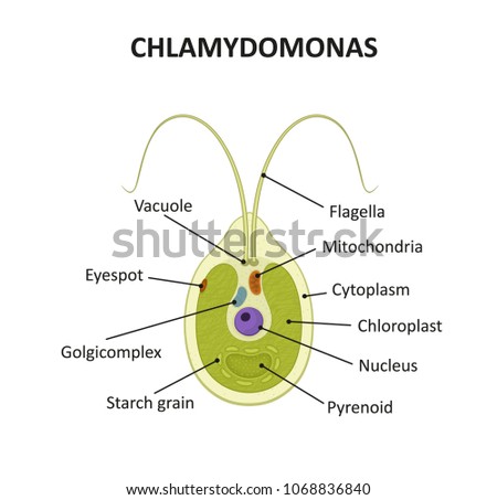



Post a Comment for "44 chlamydomonas diagram with labels"